Abstract
Triangular excision with an advancement flap is an effective yet underreported technique for pilonidal sinus disease with unilateral superolateral secondary pits, where minimally invasive methods often fail. The inverted triangular design allows complete excision of the sinus cavity and pits, and the defect is closed with a tension-free advancement flap, ensuring rapid recovery. Applicable to disease of varying vertical extent within or beyond the navicular area, this approach preserves healthy tissue and provides a practical alternative to conventional flap procedures.
Indications
The triangular excision and advancement flap is a practical option for PDS with one-sided superolateral secondary pits, particularly when minimally invasive treatments are likely to fail, and accumulated experience supports its safe and effective use while noting design considerations.,
Method
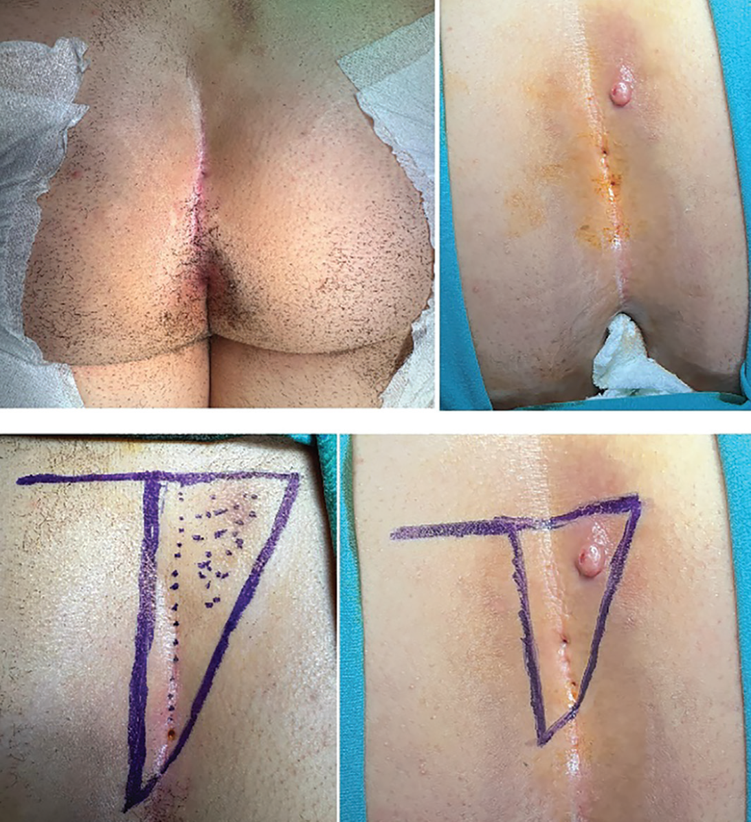
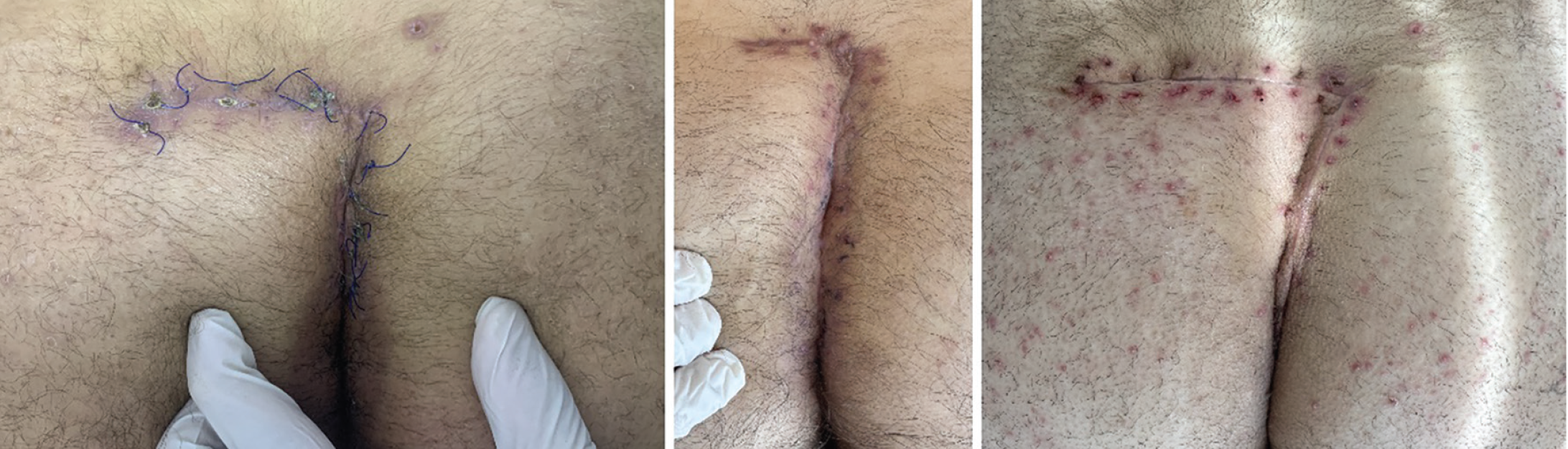
Starting with marking may be preferable. The right-angled inverted triangular design incorporates unilateral superior secondary pits, the adjacent sinus cavity, and inferior midline primary pits, with a horizontal superior base edge enabling en bloc removal of the upper affected area. The triangle’s apex, opposite the base, is located inferolateral to the most inferior pit. The vertical edge lies on the healthy side, approximately 2 cm from the midline (). Only a short segment of the hypotenuse crosses the midline, minimizing the healing challenge posed by the dynamic area. Although the vertical edge may appear midline during recovery, it lies on the “hillside” rather than the “valley bottom,” reducing exposure to intergluteal mobility, sweat, and moisture ().
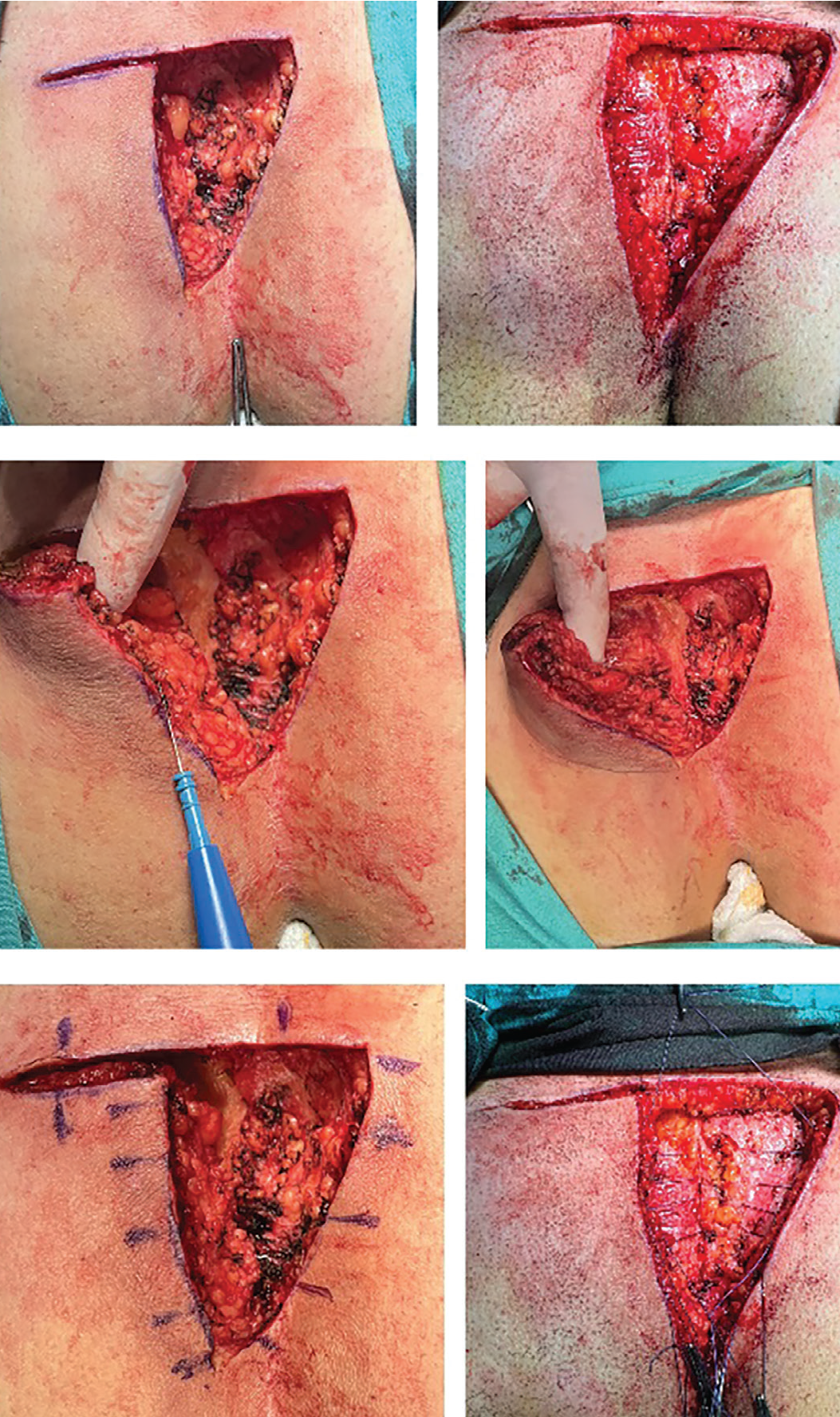
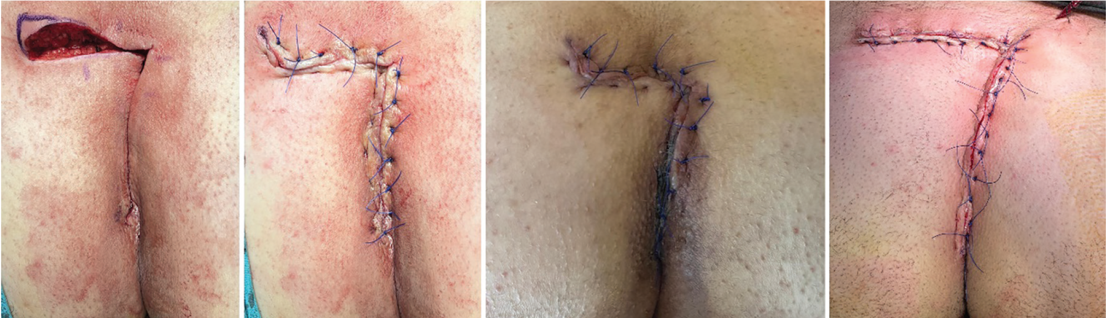
It is important to ensure that the flap is applied with minimum tension. A horizontal incision, equal to or slightly longer than the triangular excision’s base, creates a triangular peninsular fasciocutaneous flap. Meticulous dissection separates fatty tissue from the gluteus maximus muscle, and the broad base of the flap maintains vascular supply despite some perforator loss. After releasing the navicular area exposer hip tapes, flap edges are aligned and overlapped with the excision margins. The defect is closed with thick polyglactin sutures through the presacral fascia encountered after total sinus excision. In wide defects, pre-suturing placement of a closed-suction drain prevents suture interference (). This technique is applicable to pilonidal sinus disease (PDS) of varying vertical extent, including unilateral secondary pits beyond or inside the navicular area, which represent a frequently observed clinical presentation. Dog ears at the superior corner of the triangular flap base often result from extra gluteal mobilization, overlapping the apex with the opposite corner, but this can be corrected. Placement of a few sutures can draw the corner inward, allowing apex alignment with less mobilization and reduced tension, thus promoting faster healing ().
Comparison With Other Methods
In cases of superior unilateral pits extending far laterally, the Bascom cleft lift, similar to the rhomboid excision with a modified Limberg flap, requires a relatively wide excision of healthy tissue, which may prolong wound healing and increase the risk of postoperative complications., , By contrast, triangular excision with an advancement flap minimizes unnecessary tissue removal. When adequate mobilization allows for a tension-free closure, healing tends to be rapid (). Furthermore, the resulting minimal subcutaneous dead space and the total excision of omissible midline primary pits with this technique may further contribute to a lower risk of recurrence., This technique allows efficient closure and minimal sacrifice of healthy tissue, which may be advantageous in managing PDS with unilaterally extending secondary pits. In this series of 26 patients, including 4 recurrent cases amenable to this technique, all of whom provided written informed consent for publication and underwent surgery between May 2016 and September 2025, no recurrences or major early postoperative complications occurred during follow-up. These findings support previous reports suggesting that limited excision of perianal tissue may reduce the risk of perianal complications. This technical note seeks to encourage broader acceptance of this non-mainstream procedure.
Footnotes
Acknowledgments
I sincerely thank my senior colleagues at the former Okmeydanı Training and Research Hospital for introducing me to this technique, which I have had the opportunity to refine and improve over the course of more than a decade of clinical experience.
Ethics
Informed Consent: Written informed consent was obtained from all patients for publication of their data and images.
Financial Disclosure: The author declared that this study received no financial support.
References
Kızıltoprak N, Turan İ, Mazlum AF, Genç MS, Kalın M, Ercan G, Kesici U. Triangle excision and advancement flap in pilonidal disease: a single-center prospective case series. Turk J Colorectal Dis. 2024;34:69-74.
Doll D, Maak M. Refining the triangle advancement flap technique for pilonidal sinus disease: a commentary on Kiziltoprak et al. Turk J Colorectal Dis. 2024;34:145-146.
Guner A, Cekic AB, Boz A, Turkyilmaz S, Kucuktulu U. A proposed staging system for chronic symptomatic pilonidal sinus disease and results in patients treated with stage-based approach. BMC Surg. 2016;16:18.
Wysocki AP, Andersson RE, Gips M, Girgin M, Guner A, Immerman S, Kanat BH, Kayaalp C, Milone M, Petersen S, Senapati A, Tezel E, Doll D. Towards a classification for sacrococcygeal pilonidal disease – Berlin 2017. Pilonidal Sinus J. 2018;4:5-12.
Shrager B. The data mounts: 261 cleft lifts for complex pilonidal disease and excisional failures. Cureus. 2023;15:e50174.